

Images of Contents of mRNA Injections
Microscopic Images of Strange Geometric Objects In the mRNA Injections

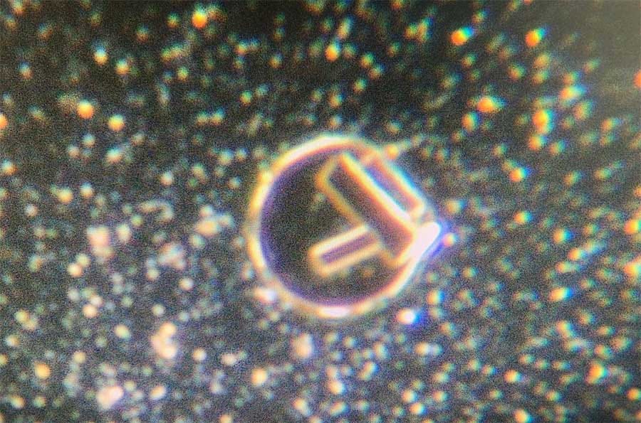
For those who just want the short story: Here’s the picture of one drop of New Zealand’s Pfizer COMIRNATY “vaccine” under a cover slip, after it was inadvertently heated lightly, and viewed later the same day through dark field microscopy at low magnification, and projected onto a TV monitor.
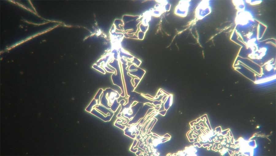
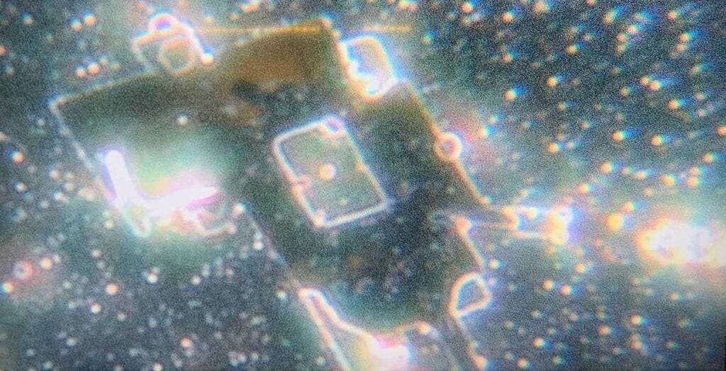
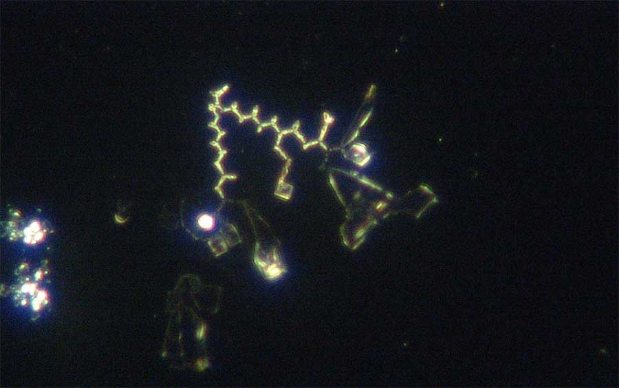
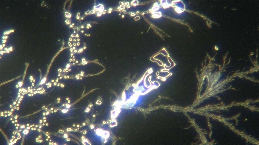
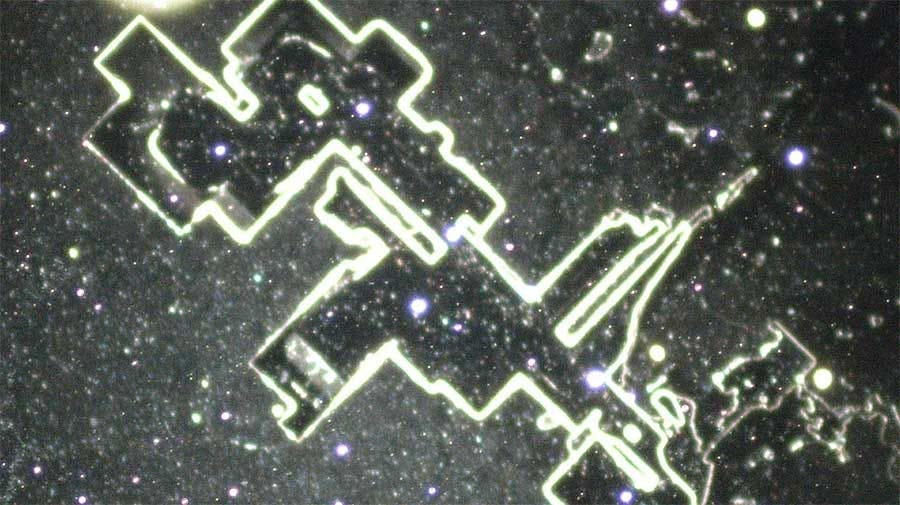
The images below are from a new sample, after a new computer with top notch graphic capacity was purchased. These images came through software connected to a microscope camera.
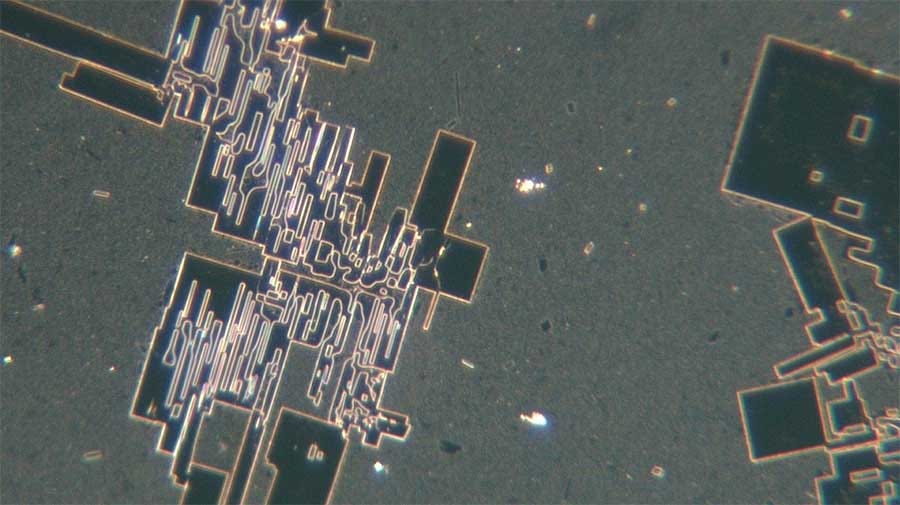
Increase the magnification on the images below and look closely
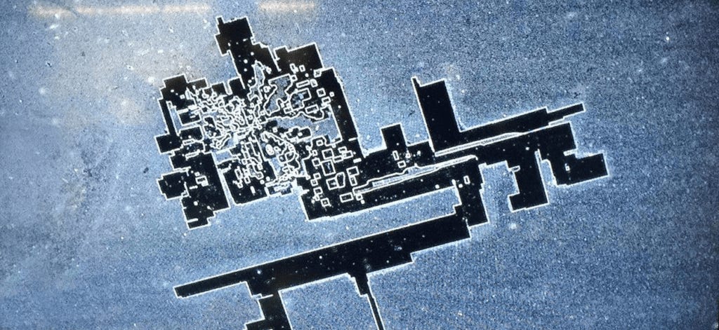
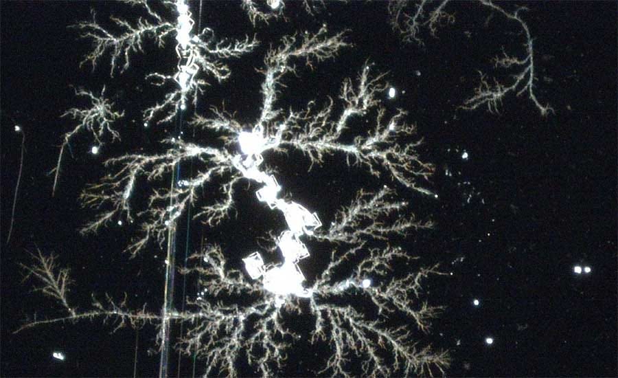


The amount of activity in the liquid and the strange shapes were baffling to say the least.
Squares and circles seemed to connect up to each other consistently. Over an hour or two, the squares and circles seemed to enlarge somehow.
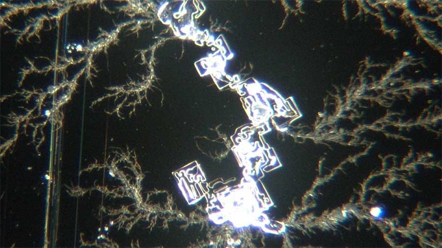
On the same hot day, I had to travel from my office to another office some distance away. The slides were packed up in plastic slide mailers and placed in a cardboard box in the car alongside the microscope. It had been a long week, and the ocean looked enticing, so I parked the car with the windows cracked about two inches, went for a 40-minute walk, then continued my drive.
At my destination, the microscope was set up and I had another look at the slides. I don’t scare easily but what I saw next gave me a bit of a chill.


The straight lines and corners were lined with squares, rectangles, and circles.
Wanting better graphics, I purchased a PC laptop with the best graphics the store could offer me. The software was then useable and I could take future images from the video program attached to the camera, for better definition.
If this was to be believed by anyone, it had to be repeated and understood better.
In the days that followed I contemplated how the formation happened. Was it a function of time? Of the vibration in the car? Heat? Cell towers along the way? It could have been any of those, so I took some more of the liquid and plated out some more slides, put them in multiple slide carriers, drove them all around the city near cell towers for two hours, and then came back.
I did get some additional interesting images but nothing like the first one.
Watch the video below FFWD to 1:18 to see this stuff in motion on microscopic videos:
https://odysee.com/@en:a5/PK_Tot-durch-Impfung_english:a
Scanning and Transmission Electron Microscopy Reveals Graphene Oxide in CoV-19 Vaccines
Abstract
Currently there are four major pharmaceutical companies who manufacture a SARS-CoV-2 now called SARS-CoV-19 vaccine. These manufactures and their vaccine are Pfizer--BioNTech mRNA Vaccine, the Moderna-Lonza mRNA-1273 Vaccine, the Serum Institute Oxford Astrazeneca Vaccine and the Janssen COVID-19 Vaccine, manufactured by Janssen Biotech Inc., a Janssen Pharmaceutical Company of Johnson and Johnson, a recombinant, replication-incompetent adenovirus type 26 expressing the SARS-CoV-2 spike protein [1]. The intended purpose of these vaccines are to provide immunity from the so-called infectious novel coronavirus or SARS-CoV-2 virus now called the SARS-CoV-19. These four pharmaceutical companies have not provided complete FDA disclosure on their vaccine box, insert fact sheet or label for many of the major and/or minor ingredients contained within these so-called vaccines.
The purpose of this research article is to identify those specific major and minor ingredients contained in the Pfizer Vaccine, the Moderna Vaccine, the Astrazeneca Vaccine and the Janssen Vaccine using various scientific anatomical, physiological and functional testing for each SARS-COV-2-19 vaccine. As a human right, governed under World Law by the Nuremberg Code of 1947, the vaccine specific ingredient information is critical, required and necessary to know so that any human from any country in the World can make an informed decision whether or not to consent to the SAR-CoV-2-19 inoculation [2]. We have conducted the scientific testing on each vaccine and have identified several ingredients or adjuvants that have not been disclosed which are contained in these four SARS-CoV-2-19 vaccines. Currently, these vaccines are being administered to millions of humans around the World under an Emergency Use Authorization (EUA) issued by each country without full disclosure of all ingredients and in some cases mandated by governments or employers in violation of individual human rights under the Nuremberg Code of 1947 [3].
Keywords: SARS; CoV-19; Vaccine; Bioweapon; 5G; Graphene; Graphene Oxide; Graphene Hydroxide; Parasite; Trypanosoma; PEG; Polyethylene Glycol; Nano Dots; rGO; GO; mRNA; Pfizer; Moderna; Astrazenica; Janssen Pharmaceutical; Electron Microscopy; Fluorescence Microscopy; Brightfield Microscopy; Darkfield Microscopy; pHase Contrast Microscopy; UV Absorbance; Fluorescence Spectroscopy; Transmission Microscopy; Energy Dispersive Spectroscopy; X-ray Diffractometer; Nuclear Magnetic Resonance; Vaccine Ingredients'.
Methodology and Techniques Four “vaccines” were analyzed which are the PfizerBioNtech, Moderna-Lonza mRNA-1273 Vaccine, Vaxzevria by Astrazeneca, Janssen by Johnson and Johnson, using different instrumentation and protocols of preparation according to new nano particulate technological approaches. The different instrumentation includes Optical Microscopy, Bright-Field Microscopy, pHase Contrast Microscopy, Dark-Field Microscopy, UV absorbance and Fluorescence Spectroscopy, Scanning Electron Microscopy, Transmission Electron Microscopy, Energy Dispersive Spectroscopy, X-ray Diffractometer, Nuclear Magnetic Resonance instruments were used to verify the “vaccines” morphologies and contents. For the high-technology measurements and the care of the investigation, all the controls were activated, and reference measurements adopted in order to obtain validated results.
Live blood phase contrast and dark-field microscopy Images of the aqueous fractions of the vaccines were subsequently obtained to visually assess the possible presence of carbon particulates or graphene. The observations under optical microscopy revealed and abundance of transparent 2D laminar objects that show great similarity with images from literature (Xu., et al. 2019), and with images obtained from rGO standard (SIGMA) (Figures 1, 2 and 3) [4]. Images of big transparent sheets of variable size and shapes were obtained, showing corrugated and flat, irregular. Smaller sheets of polygonal shapes, also similar to flakes described in literature (Xu., et al. 2019) can be revealed with pHase Contrast and Dark-Field microscopy (Figure 3) [4]. All these laminar objects were widespread in the aqueous fraction of the blood (Figure 1) or vaccine sample (Figures 2 and 3) and no component described by the registered patent can be associated with these sheets [5,6]. In figure 1 You Can See What A Cluster Bomb of Reduced Graphene Oxide (rGO) Looks Like in the Live Unstained Live Blood From the So-Called Pfizer, Moderna, Astrazeneca and Janssen “Vaccines”!
Live blood phase contrast and dark-field microscopy Images of the aqueous fractions of the vaccines were subsequently obtained to visually assess the possible presence of carbon particulates or graphene. The observations under optical microscopy revealed and abundance of transparent 2D laminar objects that show great similarity with images from literature (Xu., et al. 2019), and with images obtained from rGO standard (SIGMA) (Figures 1, 2 and 3) [4]. Images of big transparent sheets of variable size and shapes were obtained, showing corrugated and flat, irregular. Smaller sheets of polygonal shapes, also similar to flakes described in literature (Xu., et al. 2019) can be revealed with pHase Contrast and Dark-Field microscopy (Figure 3) [4]. All these laminar objects were widespread in the aqueous fraction of the blood (Figure 1) or vaccine sample (Figures 2 and 3) and no component described by the registered patent can be associated with these sheets [5,6]. In figure 1 You Can See What A Cluster Bomb of Reduced Graphene Oxide (rGO) Looks Like in the Live Unstained Live Blood From the So-Called Pfizer, Moderna, Astrazeneca and Janssen “Vaccines”!
Live blood phase contrast and dark-field microscopy Images of the aqueous fractions of the vaccines were subsequently obtained to visually assess the possible presence of carbon particulates or graphene. The observations under optical microscopy revealed and abundance of transparent 2D laminar objects that show great similarity with images from literature (Xu., et al. 2019), and with images obtained from rGO standard (SIGMA) (Figures 1, 2 and 3) [4]. Images of big transparent sheets of variable size and shapes were obtained, showing corrugated and flat, irregular. Smaller sheets of polygonal shapes, also similar to flakes described in literature (Xu., et al. 2019) can be revealed with pHase Contrast and Dark-Field microscopy (Figure 3) [4]. All these laminar objects were widespread in the aqueous fraction of the blood (Figure 1) or vaccine sample (Figures 2 and 3) and no component described by the registered patent can be associated with these sheets [5,6]. In figure 1 You Can See What A Cluster Bomb of Reduced Graphene Oxide (rGO) Looks Like in the Live Unstained Live Blood From the So-Called Pfizer, Moderna, Astrazeneca and Janssen “Vaccines”! Figure 1: Is a Micrograph of a Carbon Cluster of Reduced Graphene Oxide (rGO or Graphene Hydroxide) Viewed in the Live Unstained Human Blood with pHase Contrast Microscopy at 1500x. Note that the Red Blood Cells are Clotting in and Around the rGO Crystal in a Condition Known as Rouleau! A French Word Which Means to Chain. Dr. Robert O. Young, Profiles in Medical Microscopy, Hikari OmniPublishing, 1987 – 2021 [7,8]. Normal healthy normal blood and after mRNA Inoculation Figure 1a: Micrograph under Phase Contrast Microscopy reveals the normal healthy state of the red blood cells which are even in color, even in shape and even in size. Red Blood cells in their healthy state measure anatomically 7 microns in diameter. Dr. Robert O. Young, Profiles in Medical Microscopy, Hikari Omni Publishing, 1987-2021. Figure 1b: Micrograph taken under Phase Contrast Microscopy reveals the live blood 24 hours after the mRNA Vaccine now containing crystallized red blood cells, biological transformations of red and white blood cells, large symplasts of reduced graphene oxide or graphene hydroxide crystals center and Orotic acid crystals in the upper right hand corner of the micrograph. Dr. Robert O. Young, Hikari Omni Publishing, September, 2021 [7,8]
Nano and Micro Graphene Tubes Cause Pathological Blood Coagulation Leading to Hypercapnia, Hypoxia and Death [9]. What are the non-disclosed ingredients contained in cov-19 so-called pfizer, moderna, astrazeneca and janssen “vaccines”? To answer this question an aqueous fraction of the Pfizer, Moderna, Astrazeneca and Janssen vaccines were taken from each vial and then viewed separately under pHase Contrast Microscopy at 100x, 600x, 1000x up to1500x magnification showing anatomical evidence of reduced Graphene Oxide (rGO) or Graphene Hydroxide particulates which were compared to micrographs of rGO from Choucair., et al. 2009 for identification and verification [3]. Steps of analysis of vaccine aqueous fractions Refrigerated samples were processed under sterile conditions, using laminar flow chamber and sterilized lab ware. Steps for analyses were • Dilution in 0.9% sterile physiological saline (0.45 ml + 1.2 ml) • Polarity fractionation: 1.2 ml hexane + 120 ul of RD1 sample • Extraction of hydrophilic aqueous pHase • UV absorbance and fluorescence spectroscopy scanning • Extraction and quantification of RNA in the sample • Electron and optical microscopy of aqueous pHase. [1] The pfizer “vaccine” non-disclosed ingredients The micrographs in figures 2 and 3 were obtained using 100X, 600X, 1000x and 1500X pHase Contrast, Dark Field and Bright Field Optical Microscopy [3]. On the left of each micrograph you will view micrographs obtained from the Pfizer vaccine aqueous fraction containing rGO or Graphene Hydroxide. On the right of each micrograph, you will view a match from known sources containing GO or Graphene Hydroxide for anatomical validation. The observations under a pHase Contrast, Dark-Field, BrightField microscopy, Transmission and Scanning Electron microscopy of the vaccine product by Pfizer, including vaccine products of Moderna, Astrazeneca and Janssen revealed some entities that can be graphene materials as seen below in Figures 1 through 4. Figure 2: Shows an aqueous fraction image from Pfizer vaccine sample (left) and from reduced Graphene Oxide (rGO) standard (right) (Sigma-777684). Optical microscopy, 1000X magnification, [4,10]. Figure 2a: Is a 0.5ml aqueous fraction image from Pfizer vaccine sample viewed under pHase contrast microscopy at 1000x, showing a symplast of graphene oxide (upper left) next to a Trypanosoma cruzi parasite (lower right). Dr. Robert O. Young, Hikari Omni Publishing, September 11th, 2021 [4,10,11].
https://nouveau-monde.ca/wp-content/uploads/2022/08/rapport-robert-young.pdf
CDC Director Admits Agency Gave False Information on COVID-19 Vaccine Safety Monitoring
The director of the Centers for Disease Control and Prevention (CDC) has acknowledged publicly for the first time that the agency gave false information about its COVID-19 vaccine safety monitoring.
Dr. Rochelle Walensky, the agency’s director, said in a letter made public on Sept. 12 that the CDC did not analyze certain types of adverse event reports at all in 2021, despite the agency previously saying it started in February 2021.
“CDC performed PRR analysis between March 25, 2022, through July 31, 2022,” Walensky said. “CDC also recently addressed a previous statement made to the Epoch Times to clarify PRR were not run between February 26, 2021, to September 30, 2021.”
Walensky’s agency had promised in several documents, starting in early 2021, to perform a type of analysis called Proportional Reporting Ratio (PRR) on reports submitted to the Vaccine Adverse Event Reporting System, which it helps manage.
But the agency said in June that it did not perform PRRs. It also said that performing them was “outside th[e] agency’s purview.”
Confronted with the contradiction, Dr. John Su, a CDC official, told The Epoch Times in July that the agency started performing PRRs in February 2021 and “continues to do so to date.” But just weeks later, the CDC said Su was wrong. “CDC performed PRRs from March 25, 2022 through July 31, 2022,” a spokeswoman told The Epoch Times in August.
Walensky’s new letter, dated Sept. 2 and sent on Sept. 6 to Sen. Ron Johnson (R-Wis.), shows that Walensky is aware that her agency gave false information.
https://www.theepochtimes.com/cdc-director-admits-agency-gave-false-information-on-covid-19-vaccine-safety-monitoring_4726981.html
\\][//
Military to Trump: Apologize for Operation Warp Speed or You’re on Your Own. **
Now for those pf you who bought into the mandates to take the dangerous mRNA gene therapies, and those of you who actuall took these poisonous injections, watch now as the Official Narrative reverses itself suddeny.
https://haveitbd.com/military-to-trump-apologize-for-operation-warp-speed-or-youre-on-your-own/
\\][//